
X-RAY RUNS: Apply for Beamtime
2017 Nov 1 - Dec 21
2018 Feb 7 - Apr 3
2018 Proposal/BTR deadline: 12/1/17
2018 Apr 11 - Jun 4
2018 Proposal/BTR deadline: 2/1/18
Confocal microscopy is a powerful tool for imaging a variety of biological samples, including protein crystals. In addition to the ability to generate 3D reconstructions of the sample in question via optical sectioning, confocal microscopy can also be used to measure reaction kinetics in the fluorescence mode.
A custom-design confocal microscope was previously built by Khan et. al at MacCHESS in collaboration with Prof. Warren Zipfel of the Cornell BME department. The apparatus was capable of imaging protein crystals both in reflective and fluorescence confocal microscopy modes (Khan et al, 2012). The study showed that an off-axis confocal microscope can be used to generate “virtual” on-axis images of the protein crystals. In addition to that, the study showed that the microscope in fluorescence mode can successfully be used to locate crystals in drops that are embedded in heavy precipitate.
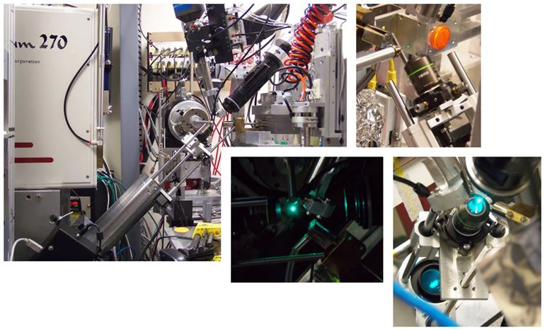
Matt Warkentin, a postdoctoral researcher from the group of Prof. Robert Thorne of Cornell University, used the apparatus at the F1 beam line to successfully measure reaction kinetics in protein crystals. A chemical reaction was triggered in the crystals by a pulse of X-rays and successfully tracked via time-lapse confocal microscopy in conjunction with x-ray diffraction imaging.
References:
[1] Khan I., Gillilan R., Kriksunov I., Williams R., Zipfel W.R., Englich U. (2012) "Confocal microscopy on the beamline: novel
three-dimensional imaging and sample positioning," J Appl Crystallogr 45(Pt 5):936-943.
Submitted by: Tiit Lukk, MacCHESS, Cornell University
01/08/2014