
X-RAY RUNS: Apply for Beamtime
2017 Nov 1 - Dec 21
2018 Feb 7 - Apr 3
2018 Proposal/BTR deadline: 12/1/17
2018 Apr 11 - Jun 4
2018 Proposal/BTR deadline: 2/1/18
Congratulations go out to the Gruner and Muller groups for their research being highlighted on the cover of the recent edition of Microscopy and Microanalysis. Their paper - “High Dynamic Range Pixel Array Detector for Scanning Transmission Electron Microscopy” - discusses the transfer of their popular x-ray detector technology into the environment of the electron microscope [1]. Pixel Array Detectors, or PADs, have brought outstanding new capabilities to x-ray instruments, such as in-pixel circuitry providing a 1,000,000:1 dynamic range within a single frame and 1.1 kHz framing rate enabling rapid data collection.
Applied to an electron microscope, the researchers discuss that by capturing the entire unsaturated diffraction pattern in scanning mode, one can simultaneously capture bright field, dark field, and phase contrast information, as well as being able to analyze the full scattering distribution, allowing true center of mass imaging. The scattering is recorded on an absolute scale, so that information such as local sample thickness can be directly determined. They describe in detail detector architecture, data acquisition system, and preliminary results from experiments with 80–200 keV electron beams.
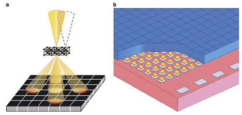
(left) Schematic of scanning transmission electron microscopy imaging using the electron microscope pixel array detector (EMPAD), and (right) schematic of the physical structure of the EMPAD (from publication, top).
The researchers conclude that while performance of this converted X-ray imager has been quite good for imaging electron scattering, some improvements could improve the utility of the device for use on electron microscopes. They propose that the real impact of this type of detector might be new data collection modalities, which will allow quantitative measures and decoupling of parameters such as tilt, electric, and magnetic fields to be extracted from a data set, or since almost every electron that is transmitted through the sample is recorded, the potential for post hoc optimally dose-efficient image reconstruction. Compared to other detectors used for diffraction inside an electron microscope, recording the full diffraction pattern on one detector allows the scattering to be placed on an absolute scale, with a direct measurement of the magnitude of diffraction spot deflections possible.
Reference:
[1] Mark W. Tate, Prafull Purohit, Darol Chamberlain, Kayla X. Nguyen, Robert Hovden, Celesta S. Chang, Pratiti Deb, Emrah Turgut, John T. Heron, Darrell G. Schlom, Daniel C. Ralph, Gregory D. Fuchs, Katherine S. Shanks, Hugh T. Philipp, David A. Muller, and Sol M. Gruner, "High Dynamic Range Pixel Array Detector for Scanning Transmission Electron Microscopy," Microscopy and Microanalysis 22 (01), 237-249 (2016).
Submitted by: Ernest Fontes, CHESS, Cornell University
03/08/2016