
X-RAY RUNS: Apply for Beamtime
2017 Nov 1 - Dec 21
2018 Feb 7 - Apr 3
2018 Proposal/BTR deadline: 12/1/17
2018 Apr 11 - Jun 4
2018 Proposal/BTR deadline: 2/1/18
K.M. Reinisha, M.L. Nibertb & S. C. Harrisona,c
aHarvard University, bDepartment of Biochemistry and Institute for
Molecular Virology, Univ. of Wisconsin-Madison, Madison, WI, cHoward
Hughes Medical Institute, Cambridge, MA
[Nature, Vol 404, 960-967 (2000).]
Reoviruses infect the respiratory and intestinal tracts of mammals and
birds. Although infections seldom cause disease, reoviruses are related
to other more serious viruses. The family reoviridae includes a
large number of double-stranded RNA viruses infecting vertebrates,
invertebrates, and plants.
Synchrotron radiation is the only tool available for the
determination of very large molecular structures at high resolution
such as the reovirus core. One of the largest structures solved
to-date has been reported from work carried out at MacCHESS by Karin
Reinish in the Harrison group at Harvard. The reovirus core is a
macromolecular assembly with a molecular mass of 52 million. The
core synthesizes, modifies, and exports viral messenger RNA. The
core contains five of the eight proteins that make up a complete
virion and is about 700 Angstroms in diameter. They crystallize in a
centered cubic space group with unit cell dimensions of 1255
Angstrom with crystal growth requiring 9 to 12 months. Using the
CHESS F1 facility, one of the two Biosafety Level 2 facilities in
the US, scientists have been able to "see" into the
three-dimensional structure of the core using the tools of x-ray
crystallography.
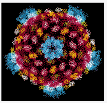
The reovirus core particle shows the subunits in different colors.
There are 120 copies of the part in red that forms the shell and
that packages the RNA. This part defines the symmetry and size of
the particle. Other subunits, shown in yellow, green and white
stabilize the shell. The blue parts form turret-like structures
around the fivefold axes that exports mature mRNA into the cytoplasm
of the infected cell.
Learning how this assembly works is one important step in learning
how to control what hazardous viruses can do to mankind.