
X-RAY RUNS: Apply for Beamtime
2017 Nov 1 - Dec 21
2018 Feb 7 - Apr 3
2018 Proposal/BTR deadline: 12/1/17
2018 Apr 11 - Jun 4
2018 Proposal/BTR deadline: 2/1/18
High-energy (HE) x-ray diffraction (energies 30 keV and above) have long been a mainstay at CHESS, supporting many user groups on a wide variety of in-house developed techniques. HE synchrotron x-rays are well suited to in-situ and operando studies of energy and engineering materials, due to their short wavelength and narrow bandpass, adjustable energy and beam size, high flux and ability to interrogate samples in complex environments by penetrating electrochemical cells or bulk polycrystalline samples up to centimeters in thickness. Pressure to grow the HE program is now coming from research groups in engineering – who need to study stress, strain, fatigue and crack formation – and chemists and materials scientists, needing to learn more about novel energy materials comprising catalysts (1) and batteries and fuel cells (2), to name a few. Investigations of these important materials and the challenges of such systems resulted in oversubscribed HE beamlines in the US and demand is continuing to grow.
In order to address this growing demand, CHESS extensively upgraded the F2 beamline and hutch over the past year to support HE experiments with intense, focused x-ray beams of energies from 40-80 keV. By partnering with the Materials Directorate of the Air Force Research Laboratory (AFRL) at Wright Patterson Air Force Base and the EMC2 group at Cornell, CHESS codeveloped a new cryo-cooled double-Laue monochromator, rebuilt a significant portion of the F-line front end, completely re-instrumented the F2 hutch, commissioned three new fast large-area detectors, designed a state-of-the-art load frame and control system, established new data acquisition hardware and software, developed software for data analysis, and procured new state-of-the-art workstations to efficiently analyze data at the beamline. The project to develop a new cryogenically-cooled double Laue x-ray monochromator to deliver those x-ray beams to the station was led by CHESS scientist Peter Ko, as discussed in prior news releases (3, 4).
The heart of the new F2 beamline capabilities at F2 is a novel load frame/positioning system coupled to two x-ray cameras, one for near-field diffraction and one for far-field diffraction, and one optical camera for digital image correlation measurements of strain. The in-situ load frame is a next generation instrument pioneered by the AFRL to support unobstructed diffraction data collection while specimens undergo 360 degree azimuthal rotation. The far-field camera utilizes a large-area General Electric flat-panel area detector having a 2048 x 2048 array of 200 micron pixels. A custom-built instrument control system synchronizes continuous rotation of the specimen with the various data acquisition systems responsible for collection of near- or far-field images (separately) and data from numerous other sensors at the beamline. One goal of the project was to create an instrument at CHESS that could provide measurement capabilities to complement initiatives underway at the Advanced Photon Source, the European Synchrotron Radiation Facility, and Petra III at HASYLAB.
While the new beamline serves a mix of experimental capabilities, a set of commissioning experiments were performed this summer (June 2014) to highlight the new capability to support in-situ High Energy Diffraction Microscopy (HEDM). HEDM experiments can address a critical need with respect to developing new materials and processing conditions by validating models and industry-standard tools focused on the prediction of mechanical properties below the continuum-scale, where the response of grains and similar microstructural features are explicitly tracked (e.g., Crystal Plasticity Finite Element Model, Field Dislocation Mechanics, etc.) (5). These validation experiments consist of in-situ loading experiments at ambient and elevated temperatures, where synchrotron x-rays are used to track the evolution of elastic strain each individual grain throughout the aggregate, as well as complementary diffraction tomography experiments to map both the morphology and local crystallography of grains within the test sample.

Grain map of Austenite measured at F2 with 55 keV. The sample cross section was 0.7 mm by 0.7 mm.
Grain mapping is a nondestructive method to acquire information about the shape, orientation, location, and relationship of grains within a bulk polycrystalline specimen. This technique is important to the structural materials community, especially in modeling applications, since it provides information about the detailed environment of neighboring grains. We are pleased to report that, during the last run cycle, the first round of commissioning experiments were successfully performed in collaboration with Jette Oddershede, Grethe Winther and Nicolai Juul from the Technical University of Denmark (DTU), Paul Shade and TJ Turner from the Air Force Research Laboratory, Joel Bernier from Lawrence Livermore National Laboratory, Mark Obstalecki from Cornell University College of Engineering, and engineer Basil Blank from PulseRay, Inc. Near-field, far-field, tomography, and digital image correlation measurements were made on samples of austenite, titanium, and copper, to produce initial grain maps like the one below for Austenite and track material deformation. Samples were slowly deformed up to high strain, while periodic far-field measurements were made to monitor the evolution of strain and orientation in the individual grains. Already, demand from potential users who were eager to conduct their own HEDM experiments at CHESS in the near future has grown.
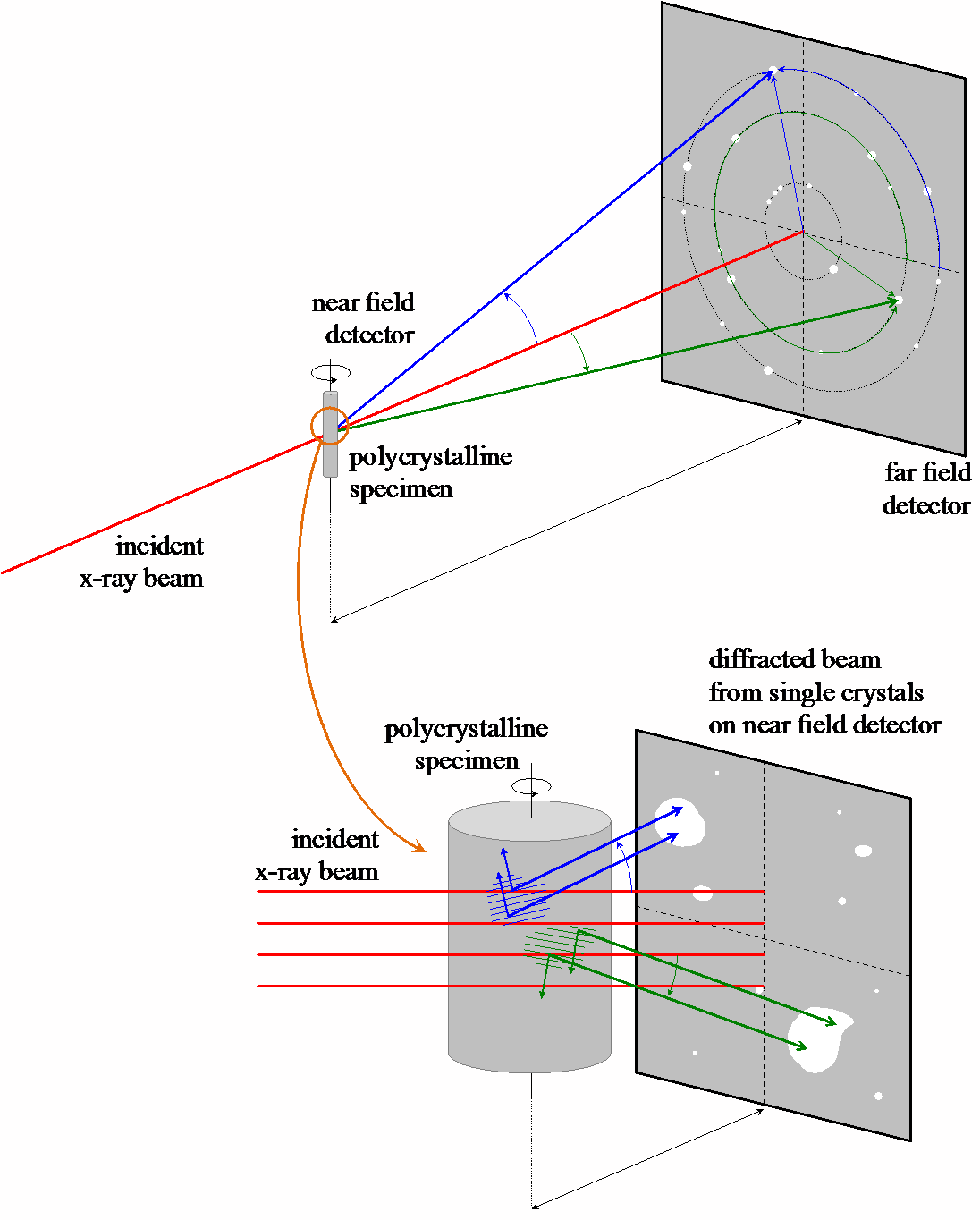
Schematic of far field and near field data collection performed on polycrystalline specimens for grain mapping techniques.
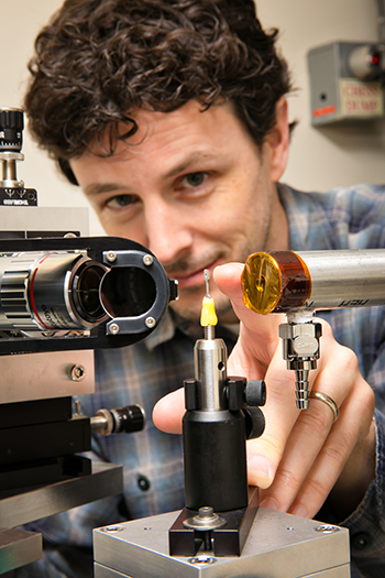
CHESS beamline scientist Darren Dale mounting test specimen during commissioning of new near-field detector (at left).
References:
[1] Gregoire JM, Dale D, Kazimirov A, DiSalvo FJ, van Dover RB, "High energy x-ray diffraction/x-ray fluorescence spectroscopy for high-throughput analysis of composition spread thin films", Review of Scientific Instruments. 2009 Dec;80(12). PubMed PMID: ISI:000273217300030. English.
[2] Liu Y, Lowe MA, Finkelstein KD, Dale DS, DiSalvo FJ, Abruna HD, "X-ray fluorescence investigation of ordered intermetallic phases as electrocatalysts towards the oxidation of small organic molecules", Chemistry. 2010 Dec 10;16(46):13689-97. PubMed PMID: 20945314. Epub 2010/10/15. eng.
[3] Fontes E, Ko JYP, "Hands-on experience makes an x-ray optics expert", 2014. Available from: http://news.chess.cornell.edu/articles/2014/Ko140312.html.
[4] Ko P, "Double-Laue Monochromator at F2", 2014. Available from: http://news.chess.cornell.edu/articles/2014/Ko140312.html.
[5] Miller MP, Suter RM, Lienert U, Beaudoin AJ, Fontes E, Almer J, et al., "High-energy Needs and Capabilities to Study Multiscale Phenomena in Crystalline Materials", Synchrotron Radiation News. 2012;25(6):18.
Submitted by: Margaret Koker, Darren Dale and Ernest Fontes
CHESS, Cornell University
08/13/2014