
X-RAY RUNS: Apply for Beamtime
2017 Nov 1 - Dec 21
2018 Feb 7 - Apr 3
2018 Proposal/BTR deadline: 12/1/17
2018 Apr 11 - Jun 4
2018 Proposal/BTR deadline: 2/1/18
Nanorods are elongated nanoparticles with aspect ratios of typically 3:1 to 10:1 and thus have interesting structural and optical anisotropies. A team of Cornell researchers led by Tobias Hanrath (Chemical Engineering) and Detlef Smilgies (CHESS) recently published a pilot study in which structure and optical adsorption were measured simultaneously, as the nanorods were exposed to solvent vapor in an in-situ cell with optical transmission ports [1]. The main goal of the study was to establish the basic relationship of the structure and optical properties of the nanorod film and how each can be influenced by external stimuli. Towards that goal, the team monitored how nanorod thin film swelled in vapor and disordered, and then re-ordered upon drying and detect the associated change in the optical properties. The phase behavior and optical properties of NRs present an interesting inorganic analogue to organic liquid crystals with potential applications in emerging optoelectronic technologies.
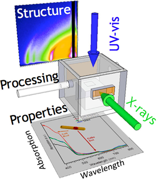
Figure 1: In-situ multi-probe set-up at CHESS D1 beamline. Grazing-icidence small- and wide-angle scattering (GISAXS and GIWAXS) as well as optical adsorption can be probed simultaneously in the in-situ cell under the same processing conditions. The in-situ cell contained dichlorobenzene in the bottom reservoir and was dried with a stream of heated nitrogen gas.
Complex dot-in-rod nanoparticles based on cadmium selenide (initial dot) and cadmium sulfide (rod) were expertly grown by Materials Science & Engineering student Christian Ocier under the supervision of Richard Robinson. Particles had a low polydispersity along and perpendicular to the rod axis with a thickness of around 5nm and lengths of 15, 20, 30, and 40 nm in the various batches of synthesis.
Both materials grow in the hexagonal wurtzite structure which provides an anisotropic diffraction pattern based on which the orientation distribution function (ODF) of the nanorods can be determined [2]. Specifically the (002) lattice planes are oriented along the long axis of the nanorods, and thus the 002 reflection acts like a searchlight indication the orientation of the rods.
By slowly evaporating the solvent from the nanorod suspension, the team was able to assemble nanorod films with long-range hexagonal ordered layers. Arrows in Figure 2a shows the characteristic 1:3:4 ratios of the scattering vectors. Wide angle scattering shows that the rod axes have a narrow orientation distribution close to perpendicular (1c). Upon exposure to solvent vapor the hexagonal layers disorder, and wide-angle scattering shows a broad distribution of orientations of the rod axes. After the layers were dried again with a preheated gas flow through the cell, they again assumed a hexagonal arrangement.
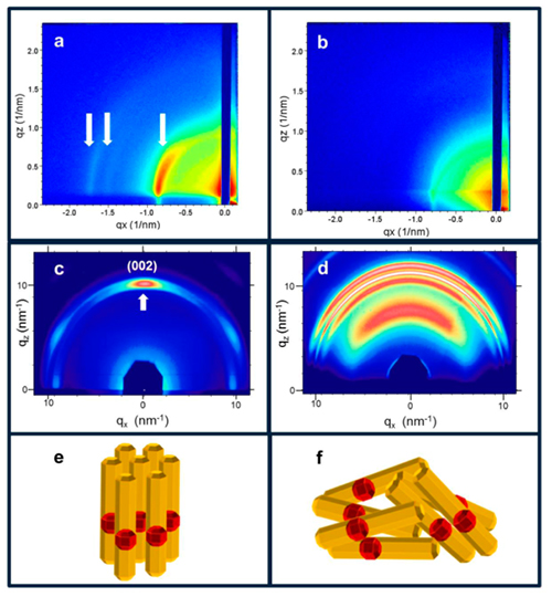
Figure 2: Grazing-incidence small- and wide-angle scattering from a nanorod layer. The layer was in the ordered hexagonal phase (a) with the rods oriented perpendicular to the surface (c) in the dry state (left column a,c,e). In the right column, the nanorods are in a disordered state (b) with their long axes featuring a wide range of orientations (d,f).
CdS and CdSe have their optical signatures in the visible range. CdSe displays an exciton peak at 600 nm. The exciton peak of CdS is shifted to the UV, however, within the spectrometer range a strong rise of the optical adsorption was visible below 500 nm wavelength. In the in-situ experiments the optical properties were probed with unpolarized white light impinging perpendicular to the glass substrate. Thus the polarization of the light was in the plane of the nanorod layer. This resulted in a significantly higher optical adsorption, when the rods were disordered in solvent vapor, than in the dry state with the rods forming a hexagonal layer with their long axes perpendicular to the substrate. Hence the optical adsorption is higher when the rod axes are oriented parallel to the surface, and thus parallel to the polarization [3]. In this work we demonstrate that multi-probe in-situ experiments can reveal the structure – optical properties relation, with a possible application to chemical sensors.
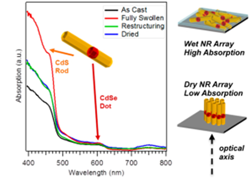
Figure 3: Optical adsorption of CdSe:CdS dot-in-rod particles. Changes in rod orientation from vertical in the ordered hexagonal lattice to parallel in the disordered phase upon solvent vapor exposure, gave rise to significant changes in the optical adsorption below 500nm wavelength.
References:
[1] Christian Ocier, Detlef-M. Smilgies, Richard Robinson, and Tobias Hanrath: "Reconfigurable Nanorod Films: An In-Situ Study of the Relationship Between Tunable Nanorod Orientation and the Optical Properties of their Self-Assembled Thin Films", Chem. Mater. 27, 2659–2665 (2015).
[2] Jessy L. Baker, Asaph Widmer-Cooper, Michael F. Toney, Phillip L. Geissler, and A. Paul Alivisatos: "Device-Scale Perpendicular Alignment of Colloidal Nanorods", Nano Lett. 10, 195-201 (2010).
[3] John Sundar Kamal, Raquel Gomes, Zeger Hens, Masoumeh Karvar, Kristiaan Neyts, Sien Compernolle, and Frank Vanhaecke: "Direct determination of absorption anisotropy in colloidal quantum rods", Phys. Rev. B 85, 035126 (2012).
Submitted by:
Detlef Smilgies, CHESS, Cornell University
06/01/2015